Sinner Research Lab
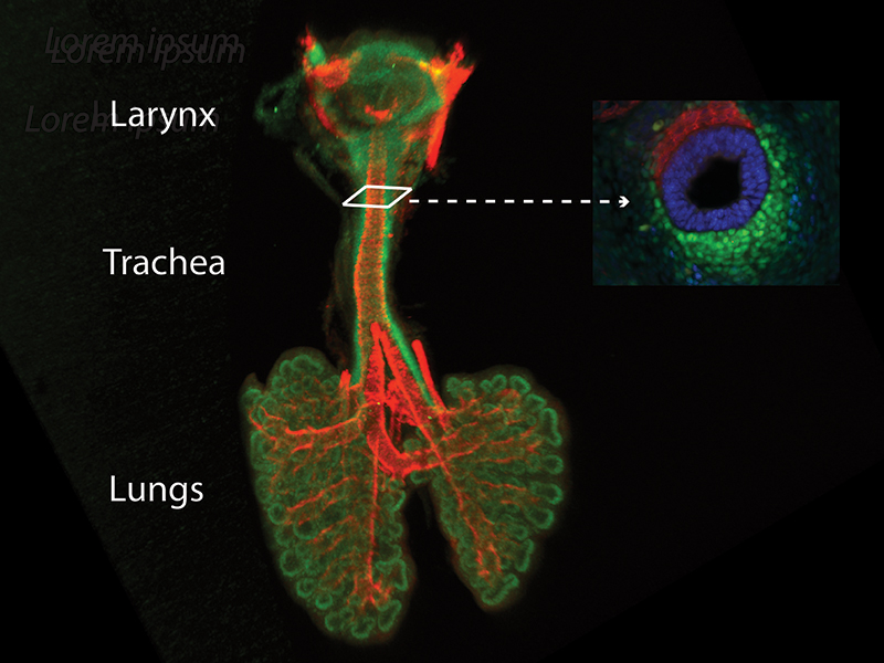
The Sinner lab aims to understand the molecular mechanisms underlying the patterning of the mammalian respiratory tract during normal development and its relevance to congenital disease. We focus on the interactions between the developing respiratory tract's two primary components: the epithelium and the mesenchyme. -The epithelium gives rise to the epithelial lining of the large airways and the respiratory surface of the lung. Cartilage, muscle, and connective tissue arise from the mesenchyme. Epithelial-Mesenchymal interactions are required for cell differentiation and are mediated by signaling pathways, including the Wnt signaling pathway that promotes the cells' differentiation and behavior during development, homeostasis, and disease.
We utilize transgenic mouse models, ex vivo culture systems, and live imaging of embryonic tracheal tissue to study the processes mediating large airways and peripheral lung formation.