Skoch Lab
Current Projects

Current Projects

Chasing Tsunamis

Spreading depolarizations or “brain tsunamis” are powerful waves of activity that spread across the cortex and are involved in a number of brain diseases including epilepsy. They are probably a fairly common occurrence but understanding their physiology and significance has been a challenge since they are very difficult to detect without invasive measures. They can be clearly seen moving across the brain with optical intrinsic signal imaging (OISI). We believe this may hold clinical potential, but we are currently studying what is referred to as the “coupling problem” to better understand how to interpret the changes seen with OISI relative to more established indicators of brain electrical activity. Using high-speed, multi-channel microscopy, we are imaging the brains of rodents to understand the spatial and temporal movement of OISI captured spreading depolarizations relative to calcium channel imaging.

An image of a brain tsunami spreading across a mouse brain.

Brain tsunami spreading across a mouse brain recorded simultaneously in 3 different optical channels.

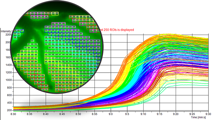
An image showing quantification of spatiotemporal progression of spreading depolarization across the brain cortex at 20x magnification.

Quantification of spatiotemporal progression of spreading depolarization across the brain cortex at 20x magnification.

Collaborators

Candi LaSarge, PhD, Steve Danzer, PhD, Matthew Batie, Carlie McCoy, Jed Hartings, PhD, and Karthik Vishwanath, PhD.

An image of a microscope.

A miniature microscope with a mount that allows rodents to be imaged.

Watching the Behaving Brain

In the field of functional neurosurgery we know that the best way to understand where seizures are coming from is to monitor them in the most “natural” way possible. This means observing patients for days or weeks with electrodes implanted in their brains and waiting for spontaneous seizure events to occur so they can be captured by our monitors. We correlate that information with videos of the patients to understand which electrical waveforms match up with certain exhibited clinical features (semiology). We hypothesize that some of the data on seizures and spreading depolarizations from slice preparations or even anesthetized in vivo models may not precisely represent spontaneous events or triggered events with anesthetic on board.

Using miniature head-mounted microscopes we aim to study seizure and spreading depolarization events in awake, behaving animals. Understanding the technical challenges of these types of recordings as well as the potential differences compared to anesthetized data is key in moving toward implementation of optical monitoring systems in clinical practice.

Collaborators

Candi LaSarge, PhD, and Steve Danzer, PhD.